
You know something is wrong. You can feel it. Maybe it is brain fog that won’t lift. Maybe it is anxiety that comes from nowhere, sleep that no longer restores you, a memory that misfires at the wrong moments, or a focus that slips through your fingers. You finally see a doctor. They order labs. They ordered an MRI. Everything comes back “normal.” And then you hear the sentence that has launched a thousand second opinions:
“We don’t know what is wrong with you.”
If you have lived this story, you are not imagining things, and you are not alone. The problem is rarely that nothing is wrong. The problem is usually that the right test has not been ordered, because the right test does not look at the brain’s structure. It looks at the brain’s function. That test is the quantitative electroencephalogram, or qEEG.
What a qEEG Actually Is
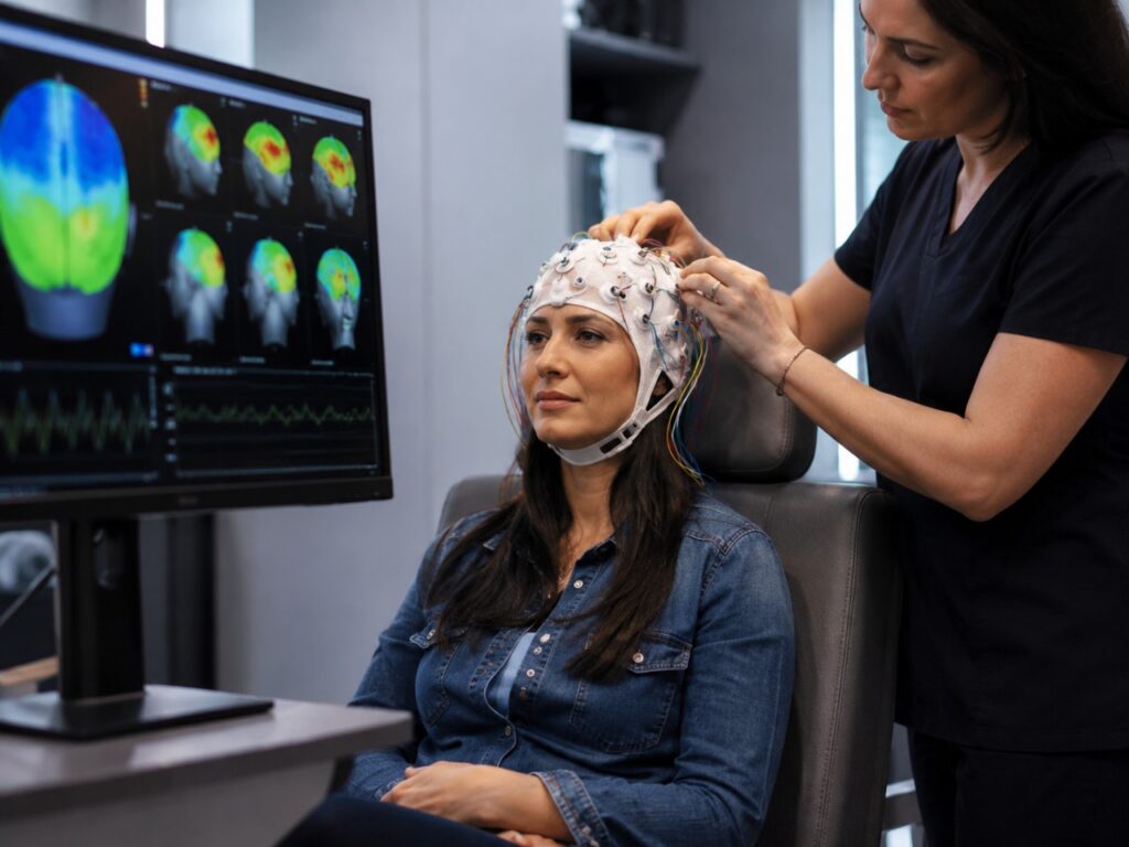
A standard EEG records the brain’s electrical activity through sensors placed on the scalp. It has been used for decades to detect seizures and gross abnormalities. A qEEG takes that same raw electrical signal and applies modern computational analysis to it. We record from 19 or more sites across the scalp while you sit quietly with eyes open, eyes closed, and sometimes while performing a simple task. The signal is then compared, frequency by frequency and region by region, to large normative databases of healthy brains matched for your age.
What emerges is a quantitative map of how your brain is working in real time — how much delta, theta, alpha, beta, and gamma activity is being generated, where it is being generated, and how different regions are communicating with one another. An MRI is a still photograph of brain anatomy. A qEEG is a movie of brain physiology. The two answer entirely different questions.
Structure Versus Function: Why the MRI Can Look Fine

MRI is exquisite at finding tumors, strokes, multiple sclerosis lesions, and atrophy. It is largely silent about dysregulation. A brain can be structurally intact and functionally chaotic. Concussion is the classic example: the MRI is almost always normal, yet the patient cannot tolerate light, cannot read for ten minutes, and cannot find a word that used to come automatically. The dysfunction is real. The MRI simply is not the instrument that detects it.
The same gap shows up in attention disorders, anxiety, and depression that resist treatment, post-COVID cognitive symptoms, sleep dysregulation, autism spectrum presentations, early cognitive decline, and the long tail of symptoms that follow viral illness, mold exposure, head trauma, or chronic stress. In all of these, a qEEG often reveals specific, reproducible electrical signatures — patterns that explain the symptoms and, just as importantly, point to what to do about them.
What the Patterns Tell Us
Excess slow-wave activity (delta and theta) in the frontal regions of an adult brain is a fingerprint we see after concussion and in attentional disorders — the brain is essentially running in a lower gear than it should. Excess high-frequency beta in certain regions is a fingerprint of anxiety and hypervigilance — the brain cannot turn the volume down. Disrupted alpha rhythms point to compromised relaxation and recovery. Coherence and connectivity measures show whether the brain’s networks are talking to each other in a coordinated way or are running as isolated islands. sLORETA source localization allows us to estimate, in three dimensions, which deeper structures — the cingulate, the insula, the precuneus — are contributing to the dysregulation.
These are not vague impressions. They are measurable, repeatable findings that explain symptoms, guide treatment, and can be tracked over time to confirm that an intervention is working.
From Diagnosis to Therapy: Neurofeedback
Once we know how the brain is misfiring, we can teach it to fire differently. That is neurofeedback. It is operant learning applied directly to brain electrical activity. The patient watches a screen, listens to audio, or plays a simple game whose output is driven by their own EEG in real time. When the brain produces a healthier pattern — less excess theta where it shouldn’t be, more alpha where alpha belongs, better network coherence — the system rewards it. Over sessions, the brain learns. The change is not pharmacologic. It is neuroplastic. And it is durable because the brain has been trained, not medicated.
Neurofeedback is most powerful when the protocol is built from the patient’s own qEEG, not from a generic template. That is the way we do it.
Where qEEG Fits in the Brain Tune Up!® Program
At Sharlin Health & Neurology, we never look at the brain in isolation. Brain Tune Up!® is built on five pillars — Move, Eat, Unwind, Discover, Connect — layered onto a functional medicine foundation that addresses genetics, methylation, inflammation, oxidative stress, glycemic control, hormones, toxins, trace minerals, and the gut. We use the Neurishment™ food guide, targeted supplementation, sleep restoration, stress regulation, and movement to rebuild the biochemical terrain the brain depends on.
qEEG enters that program in two ways. First, it is one of the most informative diagnostic studies we can run, particularly when other workups have come up empty. It tells us where to direct our attention. Second, it becomes the launching point for neurofeedback when the data show that brain training is part of what this patient needs. Foundation first, then precision.
Going Further: The Sharlin Neuro Regenerative Protocol
For some patients — those with established neurodegeneration, complex post-injury syndromes, or longstanding dysregulation that has not responded to lifestyle change alone — we go beyond the foundation into the Sharlin Neuro Regenerative Protocol. This is where qEEG-guided neurofeedback, advanced biomarker tracking, peptide and regenerative therapies, autologous stem cell approaches, and emerging longevity science come together in a single, individualized plan.
The sequence matters. We do not stack advanced therapies on a depleted, inflamed, dysregulated system and expect them to work. We build the foundation through Brain Tune Up!®, we map the brain through qEEG, and then — with a clear picture of what is broken and what is intact — we apply regenerative tools where they will actually move the needle.
Add this before the final author line:
FAQ
1. What does a qEEG show that an MRI does not?
An MRI shows the structure of the brain. A qEEG shows how the brain is functioning electrically in real time. This can help identify patterns linked to brain fog, focus issues, anxiety, sleep problems, post-concussion symptoms, and other concerns.
2. Can a brain MRI be normal even if symptoms are real?
Yes. An MRI can look normal when the issue is functional rather than structural. Symptoms such as memory lapses, poor concentration, light sensitivity, anxiety, or fatigue may still reflect measurable changes in brain activity.
3. Is qEEG brain mapping painful?
No. qEEG brain mapping is noninvasive and does not hurt. Sensors are placed on the scalp to record brain activity while you rest quietly or complete simple tasks.
4. How does qEEG guide neurofeedback?
A qEEG helps identify areas of dysregulated brain activity. That data can then be used to create a personalized neurofeedback plan that trains the brain toward healthier electrical patterns.
5. Who may benefit from qEEG brain mapping?
People with persistent brain fog, attention problems, anxiety, sleep issues, post-concussion symptoms, post-viral cognitive changes, or unexplained neurological symptoms may benefit from qEEG brain mapping.
6. Where can I schedule qEEG brain mapping in Springfield, MO?
You can schedule a consultation with Sharlin Health & Neurology in Springfield, MO, to discuss whether qEEG brain mapping may be appropriate for your symptoms and health goals.
If You Have Been Told Nothing Is Wrong
If you have heard “the MRI is normal, we don’t know what is going on,” please understand that this sentence is almost never the whole story. It usually means the test that would have shown the answer was not the one ordered. A qEEG is not exotic. It is not experimental. It is a thirty-year-old technology that has matured into one of the most useful windows we have into how a brain is working — and into what we can do to help it work better.
Your symptoms are real. Your brain is knowable. There is a path forward, and it begins with looking at the right map.
If you are searching for answers and want a deeper understanding of what may be driving your symptoms, schedule a consultation with Sharlin Health & Neurology. Our team can help determine whether qEEG brain mapping may be an important step in creating a personalized plan for your neurological health.
Kenneth Sharlin, MD
Founder, Sharlin Health & Neurology · Creator of the Brain Tune Up!® Program